Difference between revisions of "Scattering features"
KevinYager (talk | contribs) (→Spots) |
KevinYager (talk | contribs) |
||
| Line 1: | Line 1: | ||
[[Scattering]] experiments ([[x-ray]] or [[neutron]]) generates images which can be thought of as the [[Fourier transform]] of the material's [[realspace]] structure. I.e. the image is a slice through a conceptual 3D [[reciprocal-space]]. Although a scattering pattern can be arbitrarily complex, there are usually various features that can be analyzed separately, and from which one can learn much about a sample's structure. In particular, samples with well-defined structural order give rise to distinct '''scattering features''', such as sharp rings or even distinct spots on the area [[detector]] image. | [[Scattering]] experiments ([[x-ray]] or [[neutron]]) generates images which can be thought of as the [[Fourier transform]] of the material's [[realspace]] structure. I.e. the image is a slice through a conceptual 3D [[reciprocal-space]]. Although a scattering pattern can be arbitrarily complex, there are usually various features that can be analyzed separately, and from which one can learn much about a sample's structure. In particular, samples with well-defined structural order give rise to distinct '''scattering features''', such as sharp rings or even distinct spots on the area [[detector]] image. | ||
| + | |||
| + | ==Diffuse Scattering== | ||
| + | Many samples will exhibit [[diffuse scattering]]: scattering intensity over a broad range of angles, without a distinct peak or maximum. This kind of scattering usually comes from disorder within the sample. For instance, low-''q'' diffuse scattering can come from nanoscale or microscale porosity, or from surface roughness in [[GISAXS]]. High-''q'' diffuse scattering can arise from the defects in atomic lattices (and from the [[Debye-Waller factor|thermal motion]] of atoms in a lattice). Although one can generally assign diffuse scattering to some kind of disorder, it is difficult to make an unambiguous link, because there are many effects that can generate diffuse scattering. | ||
| + | |||
| + | ==Spots== | ||
| + | A sharp spot on a 2D detector is typically a [[Bragg peak]]: i.e. diffraction at a [[Bragg's law|well-defined angle]] due to a [[realspace]] [[lattice]]. An array of sharp spots typically indicates that the sample is a single-crystal (or at least [[Sample orientation|oriented]]; e.g. in-plane powder). | ||
==Rings== | ==Rings== | ||
| − | + | A ring typically indicates a [[Bragg peak]] that is spread orientationally. Thus, the sample is poly-crystalline, with crystallites at every possible orientation, but with a well-defined [[unit cell]] giving rise to diffraction spots. | |
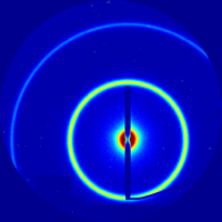
[[Image:Standard-AgBH-gisaxs th000 spot3 60sec SAXS.png|thumb|right|200px|Example [[SAXS]] with a distinct isotropic ring ([[AgBH]])]] | [[Image:Standard-AgBH-gisaxs th000 spot3 60sec SAXS.png|thumb|right|200px|Example [[SAXS]] with a distinct isotropic ring ([[AgBH]])]] | ||
| − | == | + | ==Broad Rings== |
| − | + | TBD | |
| − | == | + | ==Speckled Rings== |
| − | + | TBD | |
==Bragg Rods== | ==Bragg Rods== | ||
TBD | TBD | ||
| − | ==Yoneda Streak== | + | ==[[Yoneda]] Streak== |
TBD | TBD | ||
Revision as of 12:56, 29 January 2015
Scattering experiments (x-ray or neutron) generates images which can be thought of as the Fourier transform of the material's realspace structure. I.e. the image is a slice through a conceptual 3D reciprocal-space. Although a scattering pattern can be arbitrarily complex, there are usually various features that can be analyzed separately, and from which one can learn much about a sample's structure. In particular, samples with well-defined structural order give rise to distinct scattering features, such as sharp rings or even distinct spots on the area detector image.
Contents
Diffuse Scattering
Many samples will exhibit diffuse scattering: scattering intensity over a broad range of angles, without a distinct peak or maximum. This kind of scattering usually comes from disorder within the sample. For instance, low-q diffuse scattering can come from nanoscale or microscale porosity, or from surface roughness in GISAXS. High-q diffuse scattering can arise from the defects in atomic lattices (and from the thermal motion of atoms in a lattice). Although one can generally assign diffuse scattering to some kind of disorder, it is difficult to make an unambiguous link, because there are many effects that can generate diffuse scattering.
Spots
A sharp spot on a 2D detector is typically a Bragg peak: i.e. diffraction at a well-defined angle due to a realspace lattice. An array of sharp spots typically indicates that the sample is a single-crystal (or at least oriented; e.g. in-plane powder).
Rings
A ring typically indicates a Bragg peak that is spread orientationally. Thus, the sample is poly-crystalline, with crystallites at every possible orientation, but with a well-defined unit cell giving rise to diffraction spots.
Broad Rings
TBD
Speckled Rings
TBD
Bragg Rods
TBD
Yoneda Streak
TBD
Speckles
TBD
